Ultrasound

MicroFlow (MFI) and MicroFlow-HD (MFI-HD) imaging mode

Detect low-velocity micro-vessel anatomy in tissue without using contrast

MicroFlow Imaging (MFI) detects slow and weak blood flow anatomy in tissue. It maintains a high frame rate and excellent 2D image quality while applying artifact reduction techniques. 2D image subtraction, 2D blending and side-by-side display options offer versatility in visualization.  

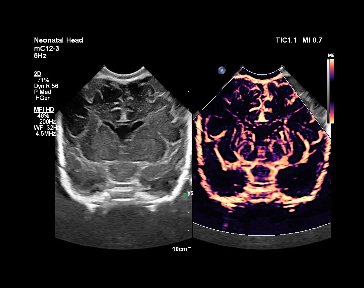
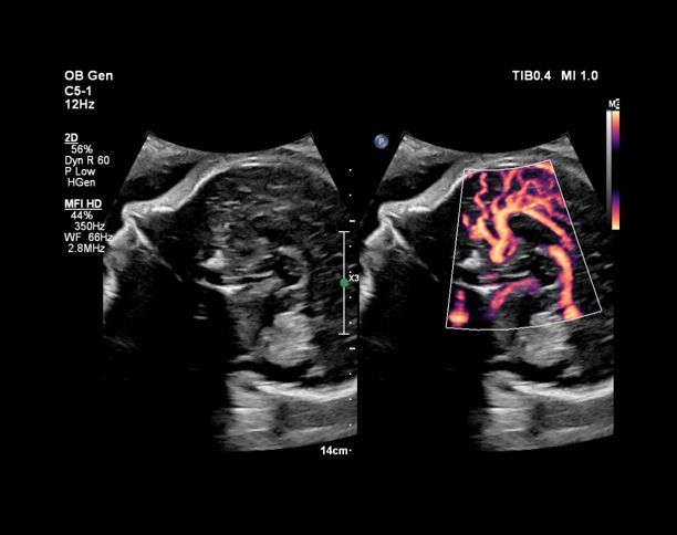
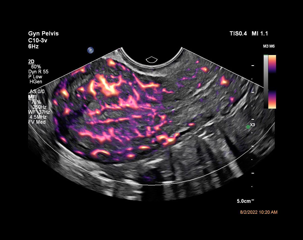
MFI-HD offers twice the sensitivity and resolution of MFI in assessing blood flow1. MFI HD is well-suited for studies requiring high resolution and sensitivity, including renal/abdominal, breast, MSK, small parts, CEUS, and OB/GYN exams.

Philips mL26-8

Featured product

MFI Image Gallery

Features

Spatial temporal clutter filter

Spatial-temporal clutter filter


MFI’s novel spatial-temporal filter analyzes Doppler data, evaluating signal characteristics from both a spatial and temporal dimension in order to distinguish the flow signals from the tissue clutter signals. In this manner, MFI can detect the lowest velocity blood flow, which microvascular vessels typically exhibit, and remove distracting tissue clutter.

Vessel enhancement algorithm

Vessel enhancement algorithm


The vessel enhancement algorithm in MFI-HD reduces noise and enhances detected vessels1 without introducing blurring often seen with more traditional image processing techniques. This vessel enhancement combines with acoustic transmits designed to optimize flow sensitivity  and spatial resolution to improve visualization with high spatial resolution.

Four display modes

Four display modes


MFI HD features four display modes based on if tissue subtraction and echo-flow compare are used, allowing customization of images. When tissue subtraction is on, MFI is focuses on displaying only the flow information, and therefore this provides the most sensitive display method. When tissue subtraction is off, MFI utilizes a tissue-flow arbitration algorithm to seamlessly blend flow with tissue information in a smooth and natural manner, therefore providing anatomical context to the visualized flow. It has the additional benefit of further tissue artifact suppression.

Ovarian blood flow with MFI

Visualize blood flow in fetal, placental, uterine and ovarian vasculature


MFI and MFI-HD help in visualizing and detecting blood flow in fetal, placental, uterine and ovarian vasculature. In a study of ovarian perfusion visualization using endocavity transducers, MFI enhanced ovarian perfusion visualization in 100% of cases.* 85% of users surveyed stated that MFI, with its ultrasensitive blood flow imaging, enhances their diagnostic confidence.**

MFI and MFI HD is available on these transducers, C5-1, C9-2, L12-3, C8-5 and eL18-4

  •  

    eL18-4 Transducer  

    The eL18-4 transducer is an ultra high-frequency linear transducer that incorporates ultra-broadband PureWave crystal technology with fine-elevation focusing capability, generating ultra-broadband frequencies from 2 to 22 MHz. The transducer’s advanced design allows for wide field-of-view trapezoid imaging and superb 2D detail resolution. It supports a broad range of high resolution applications including breast, small parts, vascular, pediatric and musculoskeletal imaging along with the penetration needed in obstetrical and gynecological examinations to help elevate clinical confidence.

    453561780782
  •  

    C5-1 Transducer  

    The C5-1 PureWave curved array transducer with 5 to 1 MHz extended operating frequency range is ideal for high performance Fetal Echo, Abdominal, Interventional, and OB/GYN applications. It offers exceptional clinical performance for a wide range of patient types including obese and technically challenging patients.

    989605412041
  •  

    C8-5 Transducer  

    Learn more about the Philips C8-5 broadband curved array transducer in the specification table below.

    989605391921
  •  

    C9-2 Transducer  

    The C9-2 PureWave curved array transducer has 9 to 2 MHz extended operating frequency range. and offers high performance for OB/GYN and Abdominal applications. Now, one transducer provides exceptional clinical performance for a wide range of patient types including technically challenging patients.

    989605391091
  •  

    L12-3 Transducer  

    Learn more about the Philips L12-3 broadband linear array transducer in the specification table below.

    989605412071

We are always interested in engaging with you.

Let us know how we can help.

1
Select your area of interest
2
Contact details

Footnotes
 

1 Internal measured comparison on standards MFI to MFI HD using clinical targets and standard measurement methodology. Not available on Affiniti systems.

*Based on a sample size of 21 users.

**Based on a sample size of 20 users.

You are about to visit a Philips global content page

Continue

You are about to visit a Philips global content page

Continue

Our site can best be viewed with the latest version of Microsoft Edge, Google Chrome or Firefox.